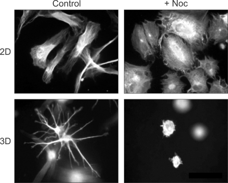
Figure 3.
Different roles of microtubules in fibroblast spreading on collagen-coated coverslips compared to collagen matrices. The actin cytoskeleton was visualized and images by fluorescence microscopy. Fibroblasts cultured for 4 h on collagen-coated coverslips (2D) spread into an elongated, flattened morphology. Disrupting microtubules with nocodazole (+Noc) inhibited cell polarization but not spreading. Fibroblasts cultured inside of collagen matrices for 4 h (3D) spread by protrusion of dendritic extension. Dendritic extensions in 3D collagen matrices were not observed in cells that were cultured with Noc. See figure 1 in (Rhee et al., 2007) for more detail.

