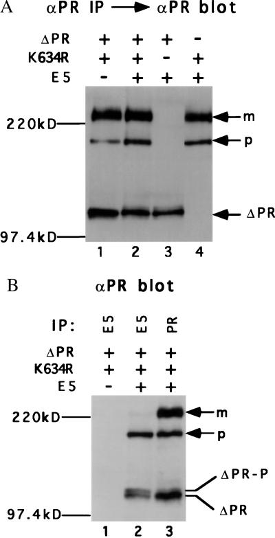
Figure 3.
Characterization of Ba/F3 cells. Extracts from Ba/F3 cells expressing the indicated proteins were immunoprecipitated with αPR (A) or the indicated antiserum (B). After gel electrophoresis, PDGF β receptor was detected by immunoblotting with αPR. The position of the mature (m) and precursor (p) forms of the full-length PDGF β receptor is shown, as is the position of the truncated receptor (ΔPR) and phosphorylated truncated receptor (ΔPR-P).

