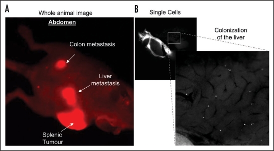
Figure 1.
(A) Whole body optical imaging of mCherry-expressing SW 620 colon cancer cell metastases after approximately six weeks post intra-splenic injection. Images were obtained using the Olympus OV100 whole body imaging system with an Olympus MT10, 150 w, Xenon light source, using a low magnification objective (macro lens) with a magnification of 0.14× and numerical aperture of 0.04. (B) mCherry expressing SW 620 colon cancer cells colonizing the liver 30 mins after intra-splenic injection. 1 × 10⁶ cells were injected into the spleen of an anesthetised CD-1 nude mouse and the incision sealed using ‘Clay Adams’ vetinary clips (VetTec). The mouse was placed on a heat pad for 30 mins then sacrificed. An incision was made in the abdomen to expose the liver and images of fluorescent cells within the liver were obtained using a 0.8× (0.22 NA) objective lens with variable zoom on the Olympus OV100.

