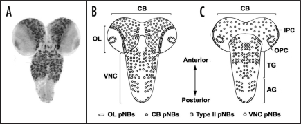
Figure 3.
The Drosophila larval CNS. (A) Overview of post-embryonic neuroblast lineages in the 3rd instar larval CNS, visualised by 1407-Gal4 driven UAS-mCD8::GFP. Schematic representation of dorsal (B) and ventral view (C) of 3rd instar larval CNS. The Drosophila larval CNS is characterised by optic lobes (OL) that consist of the inner (IPC) and outer (OPC) proliferation centres, the central brain (CB) and the ventral nerve cord (VNC) that can be sub-divided into thoracic (TG) and abdominal ganglia (AG). Postembryonic neuroblasts (pNBs) are abundantly localised in the OL, CB and VNC. A recently identified sub-group of type II pNBs are positioned within the dorso-medial region of the CB. Note that abdominal pNBs are not visualized in (A).

