
Fig. 1. Intermuscular tendons are present in the embryonic stomach.
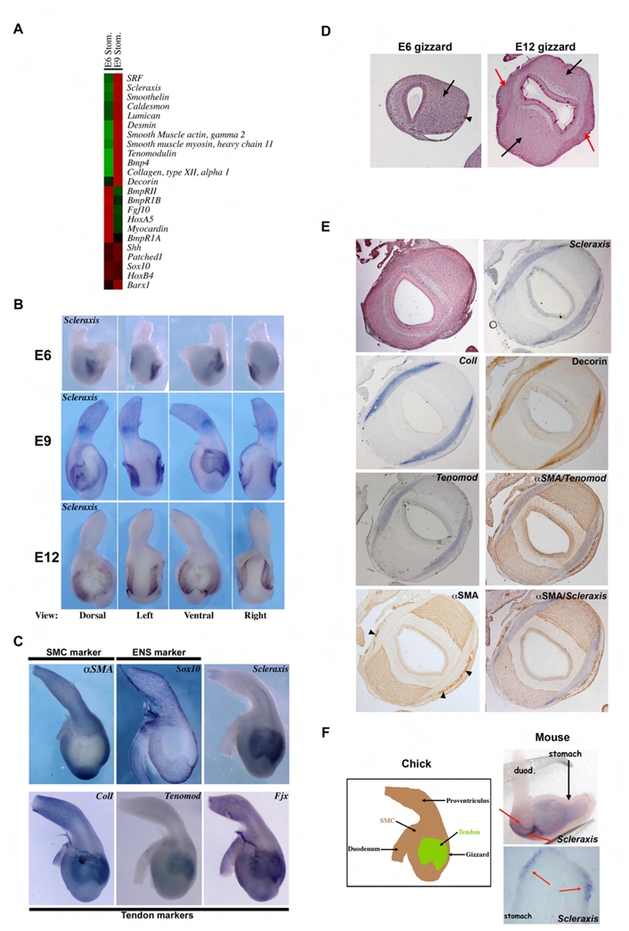
(A) Relative expression level of transcripts in E6 and E9 chick gizzards; the highest signals are in red and lowest in green. At E9, smooth muscle and tendon markers are expressed at higher levels than at E6. (B) Expression pattern of Scleraxis in the stomach by whole-mount in situ hybridization using an antisense Scleraxis riboprobe. Scleraxis expression is restricted to two newly identified domains located on the dorsal and ventral sides of the gizzard. From E6 to E9, Scleraxis expression domain widens. At E12, Scleraxis expression is restricted to the boundaries of these initial domains. (C) Whole-mount in situ hybridization on E9 stomachs. αSMA is expressed in SMC and Sox10 in ENS cells. Scleraxis, Type I Collagen (Coll), Tenomodulin and Four jointed (Fjx) are expressed mainly in the tendon domains. All tendon markers show a pattern of expression that overlaps with that of Scleraxis while they are absent from the smooth muscle domains and ENS cells. (D) Histology of E6 and E12 gizzards. At E6 the visceral mesenchyme is homogenous (arrow) in spite of the presence of migrating enteric nervous cells on the outer layer (arrowhead). At E12, the gizzard is composed of two well differentiated smooth muscle structures (black arrows) adjacent to two domains constituted of connective tissue (red arrows). (E) Serial transversal sections of an E9 gizzard. Scleraxis, Tenomodulin and Type I Collagen are detected by in situ hybridization, and αSMA and Decorin by immunostaining. Two differentiated smooth muscle structures are associated with the two emerging domains of connective tissues characterized by the specific expression of Scleraxis and of the tendon cell markers Type I Collagen, Decorin and Tenomodulin. αSMA labels the two smooth muscle areas as well as the monolayer of smooth muscle tissue surrounding the vasculature (arrowheads). In situ hybridization of Scleraxis or Tenomodulin followed by αSMA immunodetection on the same sections demonstrated the presence of two tendon structures closely associated with the visceral smooth muscle structures of the gizzard. (F) Left panel: Schematic representation of avian E9 stomach indicating the presence of intermuscular tendons. Visceral SMC domain (brown area), and well organized tendon domain (green area). Right panels: In situ hybridization on mouse E13 stomachs using a mouse Scleraxis antisense riboprobe (whole-mount and section). Two Scleraxis expression domains (red arrows) were visible.