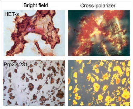
Figure 1.
Congo red binding with amyloid fibrils and bacterial inclusion bodies. Congo red staining under bright field (left) and showing birefringence under cross-polarized light (right) when binding with amyloid fibrils of HET-s (upper) and inclusion bodies of mouse prion protein PrP(23–231) (lower). (Partial reproduction of Figs. 2,26 and S8,52).

