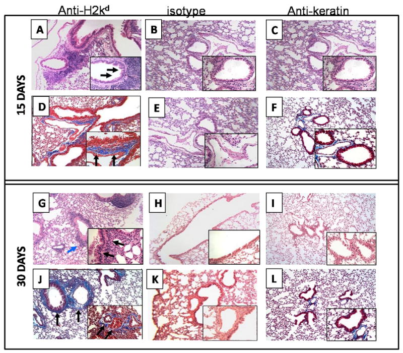
Figure 1. Administration of anti-MHC class I Abs developed significant cellular infiltration around vessel and bronchiole as well as hyperplasia of the bronchial epithelium (H&E stain).

Anti-H2kd or control (C1.18.4) Ab or anti-keratin antibody was administered endobronchially in BALB/c mice on days 1, 2, 3, 6 and weekly thereafter. The lungs were harvested on day 15 or 30 and analyzed by H&E and trichrome staining. A representative picture from data obtained from 5 mice is presented in the figure. Original magnification: × 100, insets: ×400. (A, B, C, G, H and I are sections stained with H& E; D, E, F, J, K, and L are trichrome staining. A, and D: anti-H2Kd treated mice on day 15; G and J : anti-H2Kd treated mice on day 30; B, E: C1.18.4 Ab treated mice on day 15; H, K: C1.18.4 Ab treated mice on day 30; C,F: anti-keratin Ab treated mice on day 15; I, L: anti-keratin Ab treated mice on day 30. Lungs from the anti-H2kd Ab treated mice showed significant cellular infiltration around bronchia and vessel (A) and hyperplasia of the bronchial epithelium (G) are marked by the arrows. A significant increase in the fibroproliferation, collagen deposition and luminal occlusion was observed in the lungs of the anti-H2kd Ab treated mice (J). Mice treated with isotype control Ab or anti-keratin Ab showed no significant morphological changes compared to the anti-H2Kd Ab treated mice (B, C, E, F, H, I, K and L).
