
Figure 2. Administration of anti-MHC class I Abs induces significant increase in the CD4+ and CD11b+ cells in the lungs.
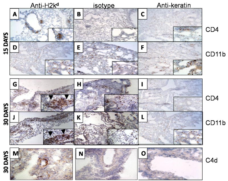
Anti-H2kd (A, D, G, J, M) or control (C1.18.4) Ab (B, E, H, K, N) or anti-keratin antibody (C, F, I, L, O) was administered endobronchially in BALB/c mice on days 1, 2, 3, 6 and weekly thereafter. The lungs were harvested on day 15 or 30 and analyzed for infiltration of CD4+ T cells, CD11b and C4d deposition by immunohistochemistry using mouse anti-CD4, anti-mouse CD11b Abs anti-mouse C4d antibodies. CD4 + cells infiltrated around the bronchiole (arrow head) and vessel at both 15 and 30 days (A, G) along with significant infiltration of CD11b + cells around the bronchiole (arrow head) and vessel at 15 and 30 days (D, J) in anti-H2kd administered mice whereas no cellular infiltration around the bronchiole and vessel was observed in isotype control (B,E, H and K) or anti-keratin antibody administered mice (C, F, I and L). A significant increase in the C4d deposition was observed in the lungs of anti-H2kd administered mice (M) when compared to control antibody administered mice (N, O). A significant increase in the levels of CD4+ T cells and CD11b+ cells was observed in the lungs of mice treated with anti-H2Kd Ab compared to mice treated with isotype control Ab.