
Figure 1.
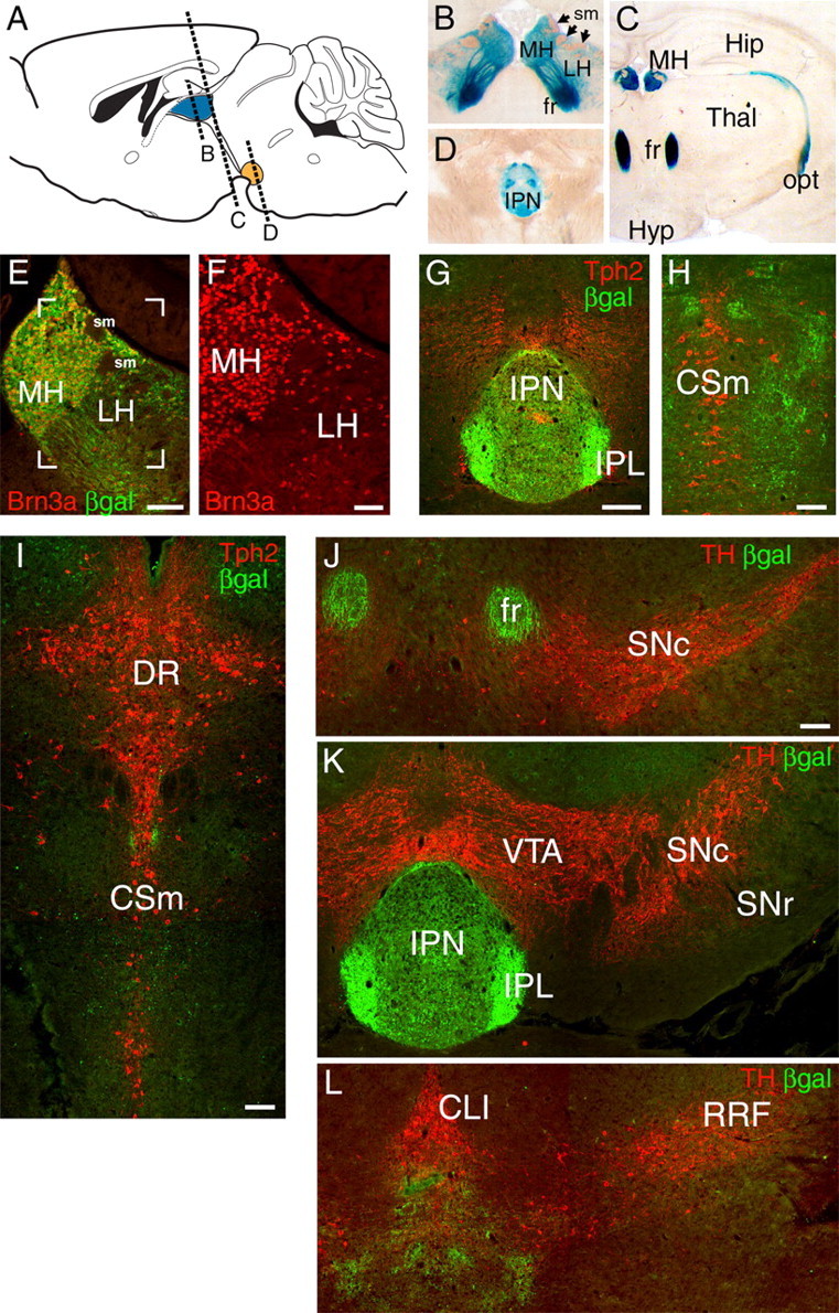
Brn3a is expressed in habenular neurons projecting specifically to the IPN. A–D, Expression of a Pou4f1tLacZ transgene in the habenula and its projections revealed by xgal staining in the adult mouse brain. Afferent habenular fibers of the striae medularis are also indicated. E, F, Colocalization of Brn3a protein expression and βgal immunoreactivity in the MH and LH. Brn3a is expressed in nearly all MH and a subset of LH neurons. The bracketed area in E is enlarged in F. G–I, Relationship of habenular projections to serotonergic neurons and fibers revealed by Tph2 immunoreactivity. G, βgal-labeled habenular fibers terminate in the IPN. Tph2 staining at this level is predominantly ascending serotonergic fiber tracts. The plane of the section is similar to D. H, I, Progressively more caudal sections showing that βgal-labeled fibers do not associate with cell bodies of serotonergic neurons of the raphe nuclei. J–L, Relationship of habenular projections to dopaminergic neurons and fibers, marked by TH immunoreactivity. βgal-labeled fibers do not project to the dopaminergic areas of the basal midbrain. CLI, Central linear nucleus raphe; CSm, superior central nucleus raphe, medial part; DR, dorsal raphe; fr, fasciculus retroflexus; Hip, hippocampus; Hyp, hypothalamus; IPL, IPN, lateral subnucleus; opt, optic tract, RRF, retrorubral field; sm, striae medularis; SNr, substantia nigra, pars reticulata; Thal, thalamus. Scale bars: E, G–L, 100 μm; F, 50 μm.