
Figure 5.
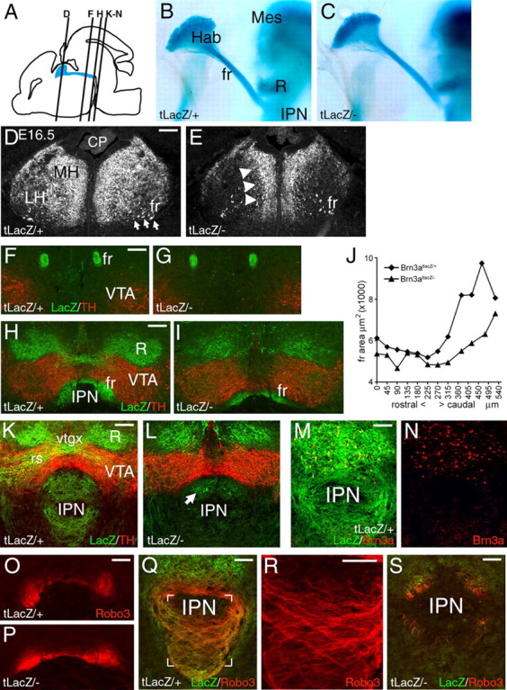
Displacement of habenular neurons and defective target innervation in Brn3a knock-out mice. A, Sagittal view of the brain at E16.5 showing the location of the habenula, FR, and IPN and the plane of sections shown in subsequent views. B, C, Midline view of habenula, FR, and IPN in hemisected, xgal-stained E16.5 embryos. D, E, Structure of the habenula in Pou4f1tLacZ/+ and Pou4f1tLacZ/− E16.5 embryos. Habenular neurons and axons are displaced toward the midline in the knock-out. F–I, Coronal sections showing the course of the FR in Pou4f1tLacZ/+ and Pou4f1tLacZ/− P0 mice, taken near the middle of its course (F, G) and just before termination in the IPN (H, I). J, Cross-sectional area of the FR from the caudal end of the habenula (zero coordinate) to the interpeduncular nucleus in P0 mice. Average values of right and left measurements are shown. The paired t test for the comparison of all sections from Pou4f1tLacZ/+ and Pou4f1tLacZ/− mice yields p = 1.0 × 10⁻⁴. K–N, Termination zone of habenular axons in the IPN at P0. The majority of βgal-immunoreactive fibers are missing in the Pou4f1tLacZ/− specimen (K, L), and the remaining expression appears to be associated with a few Brn3a+ neurons that are intrinsic to the IPN (M, N). O–S, Robo3 expression in the habenulopeduncular system. Robo3 is undiminished in the caudal FR (O, P; plane of section is similar to H). Robo3-expressing fibers innervate the IPN in a crossing pattern in a Pou4f1tLacZ/+ embryo (Q; detail R; plane of section is similar to K), but are nearly absent in a Pou4f1tLacZ/− specimen (S). CP, Choroid plexus; fr, fasciculus retroflexus; Hab, habenula; Mes, mesencephalon; R, red nucleus; rs, rubrospinal tract; vtgx, ventral tegmental decussation. Scale bars: D–I, 100 μm; K, L, O–S, 100 μm; M, N, 50 μm.