
Figure 9.
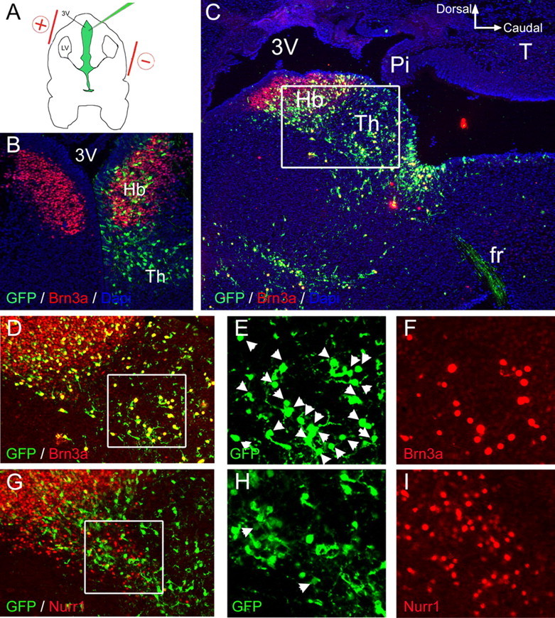
Brn3a is not sufficient to induce ectopic expression of Nurr1 in the diencephalon. A, Plasmids encoding Brn3a plus cytoplasmic or nuclear targeted GFP, or a control plasmid expressing GFP alone, were electroporated into habenular precursors by injection into the third ventricle at E13.5. Embryos were harvested at E16.5. B, Expression of GFP from a control plasmid was observed within the habenula, and in more ventral and caudal domains within the developing thalamus. The control plasmid did not alter the pattern of endogenous Brn3a expression. C, Low-power image of the habenula and periventricular thalamus in sagittal section, following electroporation of a Brn3a-GFP expression vector. Immunofluorescence for Brn3a and GFP with 4′,6-diamidino-2-phenylindole staining reveals colocalization of the expressed proteins in nearly all cells for which the nucleus resides within the plane of section. The inset box indicates the area enlarged in D and G. D–I, Expression of Brn3a (D–F) and Nurr1 in adjacent sections (G–I) after electroporation of a Brn3a-GFP plasmid. Nurr1 expression was observed in 4% of GFP+ cells in this region electroporated with Brn3a-GFP (n = 400) and 2% of cells electroporated with GFP alone (n = 400; data not shown), which did not represent a statistically significant difference. 3V, Third ventricle; Pi, pineal; fr, fasciculus retroflexus; Hb, habenula; Th, thalamus; LV, lateral ventricle.