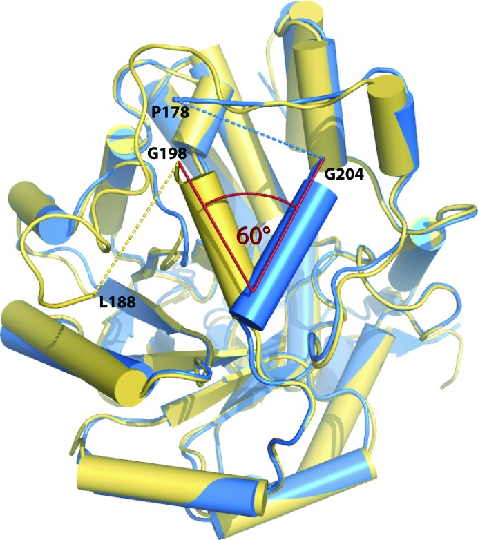
Figure 6.
Superposition of the Cα trace of an hGOX subunit (blue) with the structure of sGOX (yellow; PDB code 1gox). The disordered parts are indicated with dashed lines. The angle formed between the α-helices αE (198–206 in sGOX and 203–211 in hGOX) is indicated.

