Figure 2.
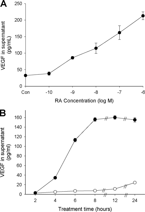
Dose response (A) and time course (B) of RA + TPA treatment on VEGF secretion from HESC. In panel A, cells were treated overnight with 50 nm TPA plus the indicated concentrations of RA. Results showed significant enhancement of VEGF secretion starting at 1 × 10⁻⁹ m RA. In panel B, cells were treated with 1 μm RA + 50 nm TPA (•) or solvent control (○) for the times indicated. Increased levels of VEGF were detectable in the supernatant starting at 4 h of treatment and peaked at 8 h. Symbols without error bars indicate that the sem was less than the value represented by the width of the symbol.

