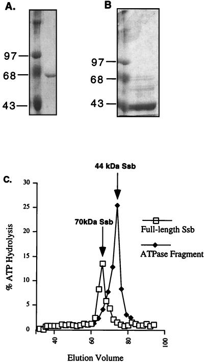
Figure 2.
Elution of Ssb ATPase domain from gel filtration column. Protein samples containing full length Ssb (A) or a spontaneously generated ATPase fragment of Ssb (B) were applied to a Sephacryl S-200 gel filtration column. In each case, the left lane is the molecular weight standard and the right lane is the protein sample. (C) Graphical representation of the coelution of full length Ssb with the ATPase activity at 66 ml (open squares) and an increased ATPase activity with the ATPase domain of Ssb at 76 ml (closed diamonds). An arrow marks the presence of each eluted protein as detected by SDS/PAGE.

