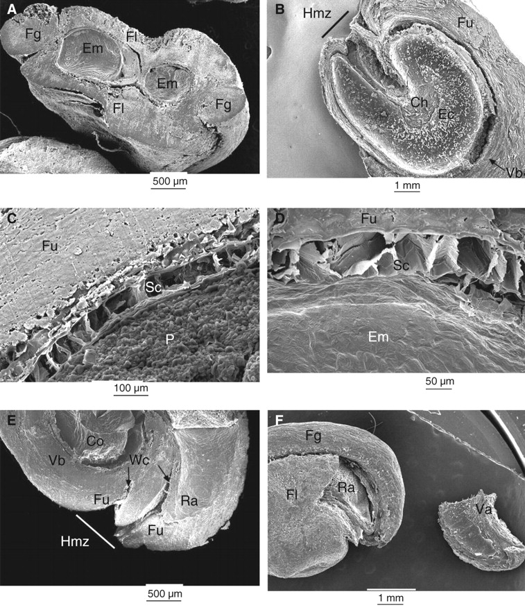
Fig. 2.

Scanning electron micrographs of (A) transverse and (B–E) longitudinal sections of O. tomentosa seeds. (B) Empty cavity in funicular envelope previously occupied by embryo. The seed coat remains can be seen attached to funiculus inside the embryo cavity. (C, D) Seed coat. (E) Hilum–micropylar zone showing water channels. (F) External morphology of seed and valve (shown on the right) detached from a dry seed. Ch, Position of the chalazal zone; Co, cotyledon; Ec, embryo cavity; Em, embryo; Fg, funicular girdle; Fl, funicular flanks; Fu, funiculus; Hmz, hilum–micropylar zone; P, perisperm; Ra, radicle; Sc, seed coat; Va, valve; Vb, vascular bundle; Wc, water channel.
