Fig. 7.
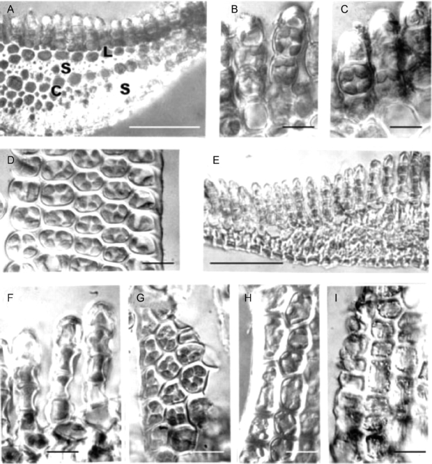
Light microscopy of Polytrichum formosum leaves. (A–D, H) Fully hydrated specimens mounted in water. (E–G, I) Completely desiccated specimens mounted in immersion oil. (A) Transverse section of a leaf midrib showing the dorsal lamellae, larger thin-walled lamina cells, stereid bands and conducting strand. (B, C) Hand-cut sections showing chloroplasts virtually filling the leaf lamella cells. No differences are apparent between the freshly collected fully hydrated specimens (B) and those hydrated from 12 h (C). (D) Surface view of a lamella. (E) In desiccated leaves the entire midrib region is highly shrunken and individual cells are difficult to distinguish. (F, G) Leaf lamella sections and surface views. Note the concave outer walls and chloroplasts the same size and shape as in fully hydrated cells. (H) Leaf lamina containing disc-shaped chloroplasts. (I) In these dehydrated lamina cells note the peripheral lipid droplets and the longitudinal striations caused by buckling of the cell walls. Scale bars: A, E=0·5 mm; B–D, F–I=10 µm.

