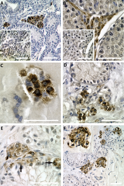
Figure 8.
Development of Leydig cells in xenografts.
Paraffin embedded sections of xenografts were stained for cytochrome p450 side-chain cleavage enzyme (p450scc, Leydig cell marker, brown precipitate), and counterstained with hematoxylin. Scale bars = 50 µm. (A, B) Positive controls. Testicular cross-sections from post-natal (A) and adult (B) rats show specific staining of Leydig cells in the interstitial compartment. Insets: negative controls. The same areas as captured in A and B in adjacent sections were incubated with secondary antibody only and do not show staining for Leydig cells. (C) Small groups of p450scc positive cells were observed in xenografts after 1 week. (D) Leydig cells become more abundant in xenografts after 4 weeks, and are located close to Sertoli cell tubules. (E, F) Progressive development of Leydig cells in long-term xenografts after 8 weeks (E) and 12 weeks (F). Most p450scc-positive cells are located in the interstitial compartment. After prolonged grafting periods, p450scc-poitive cells are also observed within Sertoli cell tubules (arrows in E).

