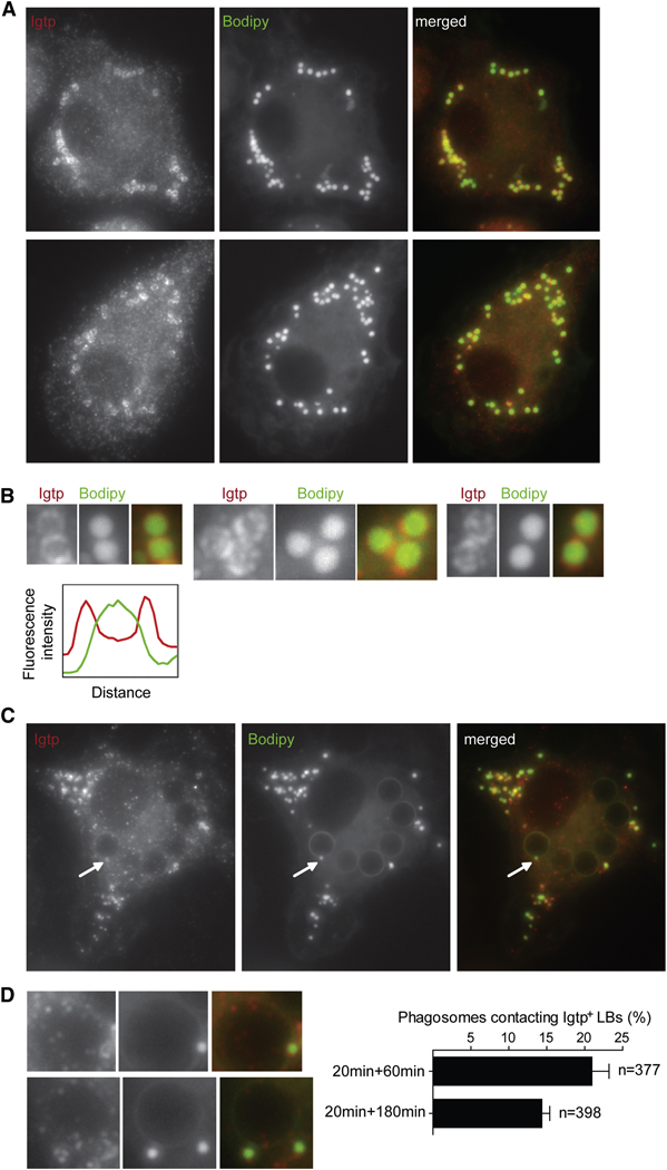
Figure 4. Irgm3 associates to LBs.
(A) Irgm3 surrounds Bodipy positive lipid bodies (LBs). IFN-γ treated WT mDCs were fixed and stained with anti-Irgm3 antibody and the LB specific probe Bodipy.
(B) Detail of Irgm3 positive LB showing the surrounding ring of Irgm3 and the dense core of the LB stained with Bodipy. The graph represents the quantification of Irgm3 (red line) and Bodipy (green line) fluorescence intensity of a representative LB.
(C) WT mDCs were fed with 3µM latex beads for 30mn, then washed and cultured for 60min. Cells were fixed and the distribution of Irgm3-positive lipid bodies was analyzed by staining as in A.
(D) Detail of LB-phagosome contact sites. A quantification of phagosomes contacting Irgm3-positive LBs at two time points after the uptake of beads is shown.

