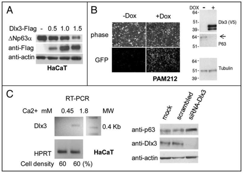
Figure 1. DlKJ reduces endogenous ΔNp63α protein in keratinocytes.

a) HaCaT cells transiently transfected with 0.5, 1.0 and 1.5 μg of Dlx3-Flag expression vector. Equal amounts of total protein (40 μg) were subjected to Western blot analysis for p63, Dlx3 (anti-Flag) and actin. b) Pictures showing OFP expression in PAM212-TetOn-V5Dlx3/GFP cells grown with (+ Dox) or without (- Dox) doxycycline for 24 hours. Phase views (upper panel) are shown to visualize cell density. Western blot of 20 μg of PAM212 nuclear extracts + and - Dox revealed with antibodies against Dlx3, p63 and tubulin. c) (Left panel) Total RNA was prepared from HaCaT cells seeded at 60% confluency (2.3 × 10⁵) in 60 mM dishes and cultured for 16 hours in 0.45 or 1.8 mM Ca2+. Dlx3 mRNA levels were determined by RT-PCR with primers specific for human Dlx3 mRNA. Values were normalized for HPRT (Hypoxanthine-guanine phospporibosyl transferase) mRNA level. (Right panel) HaCaT cells seeded at 60% confluency in 1.8 mM Ca2+ were transfected with scrambled oligonucleotides or Dlx3 siRNA) NA oligonucleotides (10 nM). 24 hours later, cells extracts were analyzed by Western blot using antibodies against p63 and Dlx3. Actin was used as loading control. MW molecular weight marker.
