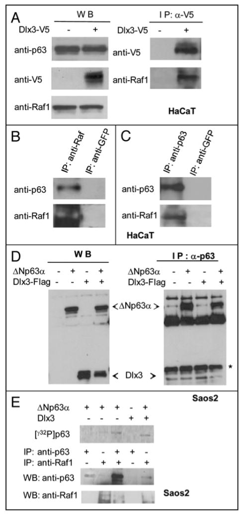
FIGURE 9. Raf-1 interacts with Dlx3 and ΔNp63α.

A) Western blot of HaCaT cells transiently transfected with V5-Dlx3 encoding plasmid and treated with 10 μM ALLNL. Equal amounts of total protein extracts from cells transfected or not were immunoblotted with anti-p63, anti-Raf and anti-V5 (left panel). Extracts from mock or V5-Dlx3 transfected cells were immunoprecipitated with anti-V5 antibody and the immunocomplexes were blotted and probed with anti-V5 or anti-Raf1 antibodies (right panel). b) Untransfected HaCaT cell lysates were immunoprecipitated with anti-Raf1 or anti-GFP antibodies. The immunocomplexes were blotted and probed with anti-p63 and anti-Raf1 antibodies. Immunoprecipitation with anti-GFP was performed as control. c) Untransfected HaCaT cell lysates were immunoprecipitated with anti-p63 or anti-GFP antibodies. The immunocomplexes were blotted and probed with anti-Raf1 and anti-p63 antibodies. d) Saos2 cells were transfected with 1 μg of ΔNp63α plasmid alone or together with 1 μg of Dlx3 plasmid. Cell extracts were immunoprecipitated with 2μg of anti-p63 antibodies. Dlx3 was revealed with antibodies to Flag. (*) Unspecific signal. e) Saos2 cells were transfected with p63 and Dlx3 vectors as indicated. Cells were treated with ALLNL. At 24 hrs extracts were immunoprecipitated with the indicated antibodies and equal aliquots incubated with 50 μM ATP and 5 μCi/sample of γ32P-ATP in kinase buffer (see materials and methods). Radiolabelled γ32P p63 (upper panel) was revealed using the PMI imaging system (BioRad). The lower panels show the immunoprecipitated p63 and Raf1 proteins as analyzed by Western blot followed by ECL detection.
