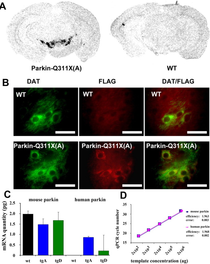
Figure 2.

Selective expression of transgene in DA neurons. A, Representative photomicrograph of in situ hybridizations using 35S-labeled parkin-Q311X antisense RNA probe on midbrain sections from transgenic mice (line A) and wild-type mice at 3 months of age. B, DAT and FLAG double immunofluorescence. Midbrain sections from Parkin-Q311X(A) and wild-type mice were immunostained using DAT (green) and FLAG (red) antibodies. Scale bar, 12.5 μm. C, D, qPCR analyses of parkin expression in laser-capture microdissected dopaminergic neurons of the SNc. Error bars indicate SEM. C, Absolute quantification of human and mouse parkin mRNA levels in WT, transgenic line A (tgA), and line D (tgD) animals. mRNA quantities are given in picograms. Note that there is no human parkin PCR product in WT animals. D, The amplification efficiencies for the mouse and human parkin cDNAs, calculated as increase in PCR product for each PCR cycle, are virtually identical. Both values are close to the theoretical value of 2.0.
