
Figure 4.
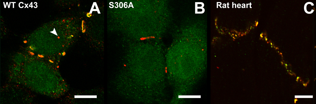
Immunofluorescence images showing total Cx43 (in red) and Cx43 phosphorylated at serine 306 (in green) in HeLa cells (A &B) and rat heart ventricular tissue (C). (A) HeLa cells transfected with WT-Cx43 display co-localization (in yellow) of the two antibodies mainly in gap junction plaques. Discrete spots only stain for total Cx43 (e.g. arrow) indicating that not all Cx43 is phosphorylated at S306. (B) S306A is not recognized by pS306 Ab in HeLa cells and therefore only total Cx43 is stained. (C) Intercalated discs in rat ventricular heart tissue are recognized by both antibodies showing that most of this Cx43 is phosphorylated at S306. Scalebars are 10 µm.