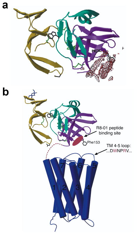
Fig. 2.
Structure of the Mth ectodomain in complex with the R8-01 15-mer peptide. (a) Electron density reveals the putative peptide binding site on the Mth ectodomain (shown as a ribbon diagram) from an averaged 3.5 Å FO – FC map contoured at 9 σ. Trp120 (a previously proposed natural ligand binding site9), Tyr130 (at the R8-01 binding site), and Asp46 and Phe153 (suggested to interact with the extracellular face of the TM domain9) are shown as stick models. (b) Scaled model depicting the full-length structure of Mth (adapted from 9). The TM domain is depicted by the structure of rhodopsin30 as a representative GPCR.

