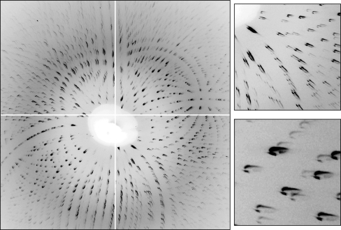
Figure 3.
Laue diffraction image taken from a small lysozyme crystal, with magnified views of two small regions of the image. The divergence from the capillary was too large, resulting in many overlapping diffraction spots. The odd shapes seen in these particular spots represent approximately half of the full far-field pattern (Fig. 5 ▶), with a small portion of the direct beam (owing to pre-capillary beamstop misalignment). If the full divergence from the capillary were used, the diffraction spots would have elliptical shapes.

