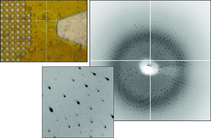
Figure 4.
Laue diffraction from a lysozyme crystal. Left: crystal on a MicroMesh mount. The circle around the crystal is ∼100 µm in diameter; the crystal is about 30 µm across. Right: diffraction pattern from the crystal with a 10 s exposure time. The inset shows well separated acceptably shaped spots.

