Figure 4.
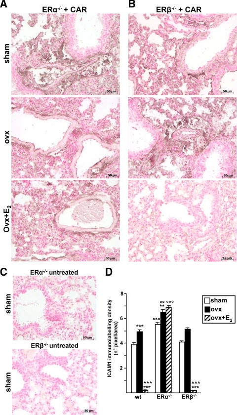
ICAM-1 immunolabeling in lung of ER-KO animals. Lung tissue from ERα- (A) or ERβ-KO (B) mice, sham, ovx, or ovx+E2, were analyzed by IHC after CAR injection with specific antibody against ICAM-1. C, IHC of lung sections from ERα- and ERβ-KO untreated mice. Representative immunhistochemistry images are shown. Densitometry evaluation (D) of ICAM-1 immunolabeling in wt, ERα-KO, and ERβ-KO mice. Bar legend as in Fig. 1B. Data are expressed as means of percent of total tissue area ± sem. * vs. sham, ^ vs. ovx; ° vs. corresponding treatment in wt. **, °°, P < 0.01; ***, °°°, P < 0.001.

