Figure 2.
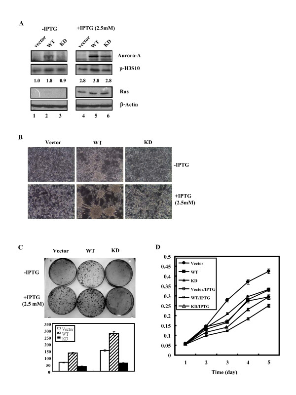
Stable overexpression of wild-type or kinase-dead Aurora-A in WT and KD cells. (A) Expression levels of human Aurora-A protein (wild-type: WT; kinase-inactivated: KD) and Ras in the Vector, WT and KD cells were detected by western blot analysis using anti-Aurora-A and anti-Ras specific antibodies. β-actin was used as the equal loading control. The levels of p-H3S10 protein were quantified by a densitometer. The expression level of p-H3S10 in Vector cells without IPTG induction was set as 1.0 fold. (B) Morphology of Vector, WT and KD cells with or without IPTG induction. (C) Upper panel: Cells (5 × 10²) were grown in 10 cm culture dishes with or without IPTG inductionfor two weeks. The foci were stained using Giemsa stain. Lower panel: Quantitative foci numbers of Vector, WT and KD cells with or without IPTG induction. (D) Growth curve of cell lines was measured daily for 5 days using MTT assay. The experiments were conducted in triplicate and repeated three times.

