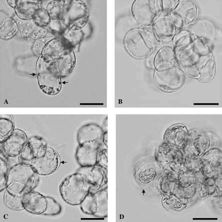
Fig. 1.
Morphology of living and AL-PCD cells from A. thaliana cell suspension cultures. (A, B) Living cells from (A) light-grown culture and (B) dark-grown culture. Note the large organelles in the light-grown cells (A), indicated by the two arrows, which were absent from the dark-grown cells (B). (C, D) Dead cells from (C) light-grown culture and (D) dark-grown culture, 24 h after heat treatment. These cells displayed the AL-PCD hallmark morphology of cell condensation, visible as a gap between the cell wall and cytoplasm, indicated by the arrows in both (C) and (D). Note that the cell condensation was far more extreme in dark-grown (D) than in light-grown (C) cells. Scale bars represent 20 μm.

