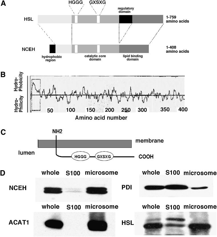
Fig. 1.
Subcellular distribution of NCEH. A: Structure of HSL and NCEH. B: A hydropathy plot of NCEH produced with the SOSUI program. C: The membrane topology of NCEH predicted by the TMHMM program. D: MPMs fractionated as described in Materials and Methods. S100 and microsomal fractions were prepared by centrifugation of the sonicated MPMs at 100,000 g for 45 min at 4°C twice. Aliquots (10 µg) of whole-cell lysate, the S100, and microsomal fractions were analyzed by Western blotting with anti-NCEH, HSL, ACAT1, and PDI antibodies. Data are representative of three independent experiments.

