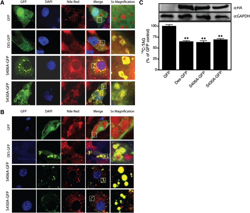
Fig. 4.
Phosphorylation of desnutrin at S406 and S430 is not essential for activity or localization. Confocal micrographs showing diffuse localization of GFP (control) or prominent lipid droplet localization of GFP-tagged desnutrin, S406A, and S430A desnutrin mutants in COS-7 cells (A) or 3T3-L1CARΔ adipocytes (B). TAG hydrolysis by desnutrin-GFP or serine to alanine mutants of desnutrin-GFP measured in live 293FT cells (C). Immunoblots show expression of desnutrin and mutant desnutrin and GAPDH (control). Values are means ± SEM from a representative experiment. **P < 0.01 versus GFP control.

