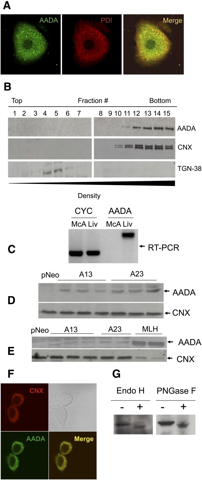
Fig. 1.
Localization and expression of mouse AADA. A: Localization of endogenous AADA in primary mouse hepatocytes by confocal immunofluorescence microscopy. B: Subcellular fractionation of mouse liver homogenate. AADA was detected with anti-mouse AADA polyclonal antibodies as described in Materials and Methods. C: AADA is expressed in rat liver but not in rat-derived McA cells as determined by RT-PCR. CYC, cyclophilin (control); Liv, liver. D: Twenty micrograms of membrane proteins prepared from individual dishes of McA cells stably transfected with an empty pCI-neo vector or pCI-neo vector containing FLAG-tagged mouse AADA cDNA (A13 and A23) were electrophoresed in SDS-PAGE, proteins were transferred to a nitrocellulose membrane and immunoblotted with anti-FLAG and anti-CNX (loading control) antibodies as described in Materials and Methods. E: Twenty micrograms of membrane protein from stably transfected McA cells and from mouse liver homogenate (MLH) were electrophoresed, transferred to a nitrocellulose membrane, and immunoblotted with anti-mouse AADA and anti-CNX antibodies as described in Materials and Methods. F: Localization of AADA in transfected McA cells by confocal immunofluorescence microscopy. G: Cell lysates from AADA-expressing cells were treated with deglycosylation enzymes and changes in AADA molecular mass was assessed by SDS-PAGE.

