Fig. 4.
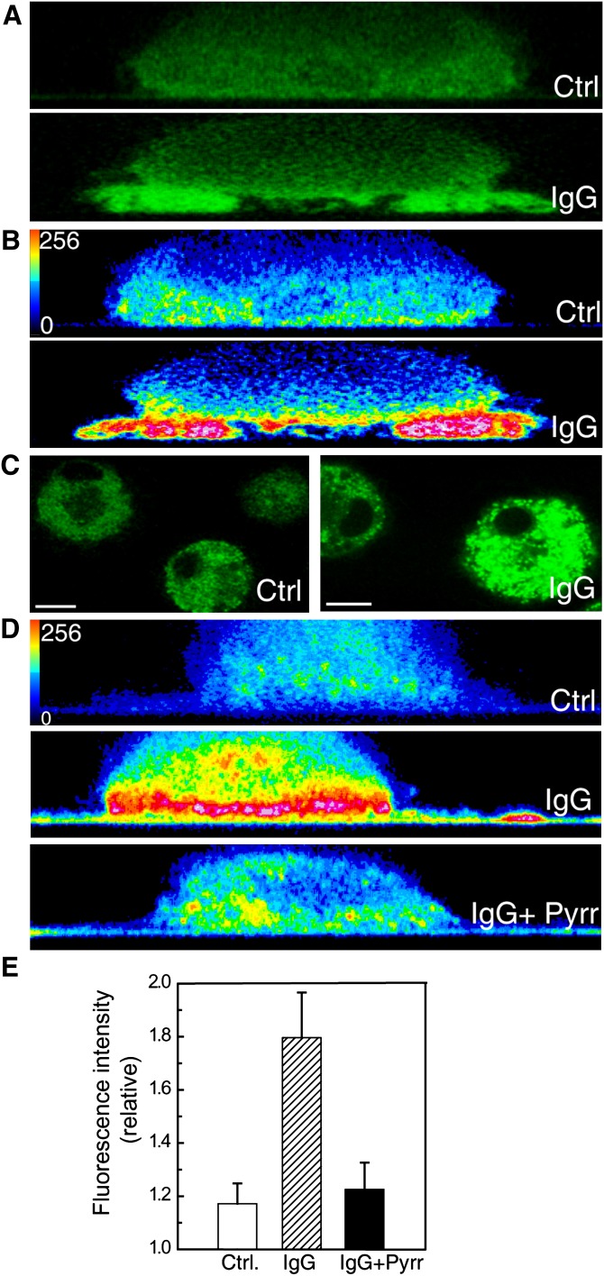
cPLA2α is functionally active in the phagosomal cup in human macrophages. A: Cells transfected with EGFP-cPLA2 were subjected to a frustrated phagocytosis assay on noncoated glass (Ctrl) or IgG-coated glass (IgG), as indicated, and analyzed by confocal microscopy. Pictures of the XZ axis of the cells were taken. B: The intensity of the fluorescence obtained in (A) was analyzed in pseudocolor. C: Cells, labeled with bis-BODIPY FL C11-PC, were plated on noncoated glass (Ctrl) or on IgG-coated glass (IgG) for 30 min, and fluorescence was analyzed by confocal microscopy. The mean of fluorescence intensity in Ctrl was 66 and in IgG was 128. D: The cells were labeled with bis-BODIPY FL C11-PC and subjected to a frustrated phagocytosis assay. Pictures of the XZ axis of the cells were taken and the intensity of the fluorescence was analyzed in pseudocolor. In the picture on the bottom, the cells were treated with 1 μM pyrrophenone (Pyrr). White bar = 10 μm. E: Statistical analysis of the fluorescence intensity in cells assayed for frustrated phagocytosis as in D. Data are represented as relative fluorescence intensities (mean fluorescence intensity in membranes closer to the glass/mean fluorescence intensity in the cytosol). At least 15 different cells were analyzed.

