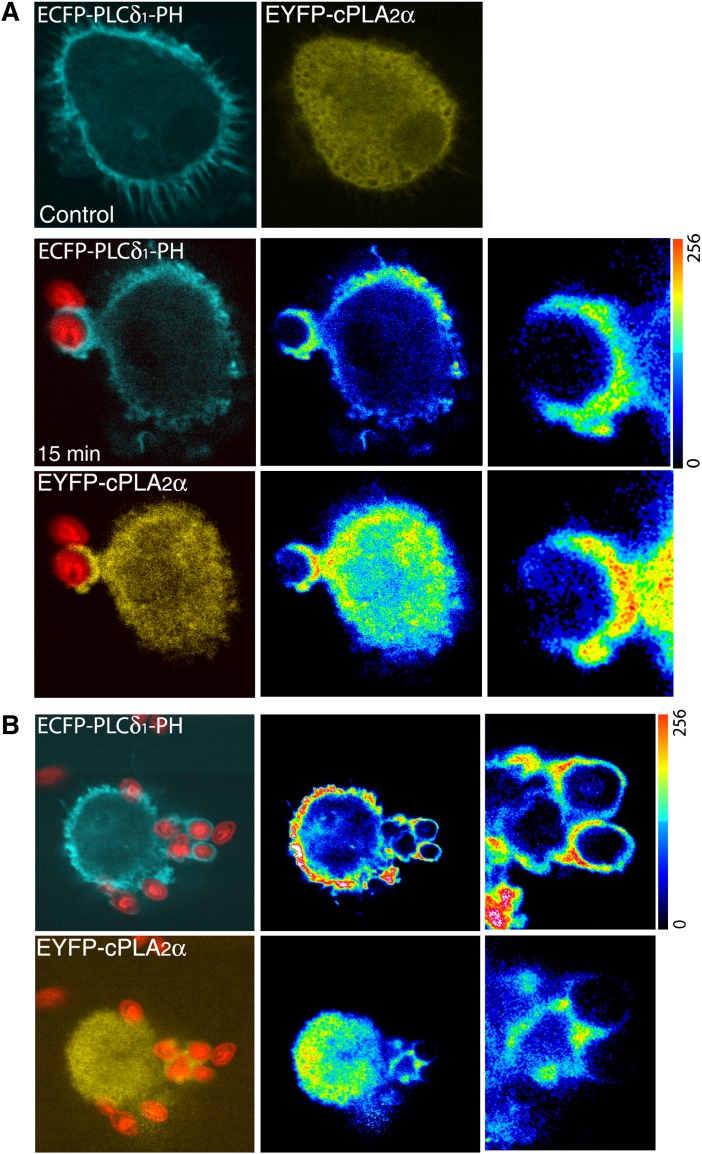
Fig. 7.
Localization of ECFP-PLCδ1-PH and EYFP-cPLA2α during phagocytosis in human macrophages. Human macrophages, cotransfected with the ECFP-PLCδ1-PH and the EYFP-cPLA2α constructs, were subjected to synchronized phagocytosis using Alexa Fluor 594-labeled opsonized zymosan, fixed at 0 (Control) and 15 min, and analyzed by confocal microscopy (A). B: A cell with high expression of the construct ECFP-PLCδ1-PH is shown. In A and B, middle panels, fluorescence intensities are shown in pseudocolor, and detailed fluorescence in forming phagosomes is shown in panels to the right.

