
Figure 3.
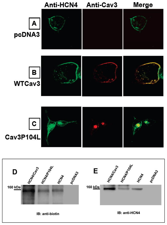
Confocal images from HCN4 stably expressed HEK293 cells transiently expressed with pcDNA3 (A), WTCav3 (B), or Cav3P104L(C). The cells were immunostained with rabbit anti-HCN4 polyclonal antibody and mouse anti-Cav3 monoclonal antibody. Cell surface biotinylation of HCN4 channels was examined in HEK293 cells transiently transfected with HCN4, HCN4/Cav3, HCN4/P104L, or pcDNA3. Biotin-labeled HEK293 cell lysate was immunoprecipitated with anti-HCN4 antibody and subjected to Western blot analysis with antibiotin antibody (D), then stripped and reprobed with anti-HCN4 antibody (E); the 160 kDa band for HCN4 is indicated.