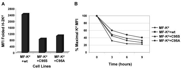
Figure 4.
Stable surface expression of Kd was impaired by mouse tapasin C95S and tapasin C95A compared to wild type tapasin. (A) Relative to cells expressing wild type mouse tapasin, cells expressing mouse C95S tapasin or C95A tapasin had a reduced level of cell surface Kd. Cells were incubated with an antibody against the folded form of the Kd molecule (34-1-2) or with secondary antibody only. Results obtained with the secondary antibody only were less than 4.0. Values on the y axis are relative mean fluorescence intensity (MFI) units obtained with antibody 34-1-2. (B) Surface Kd molecules on cells expressing mouse tapasin C95S or tapasin C95A had a faster turnover rate than those assembled in the presence of wild type mouse tapasin. MF cells expressing Kd with no tapasin, wild type tapasin, tapasin C95S, or tapasin C95A were incubated with 2 μg/ml brefeldin A in complete medium for 0, 3, 6, or 9 hours. After the incubation with brefeldin A, the cells were washed, stained with anti-Kd antibody 34-1-2, and analyzed by flow cytometry.

