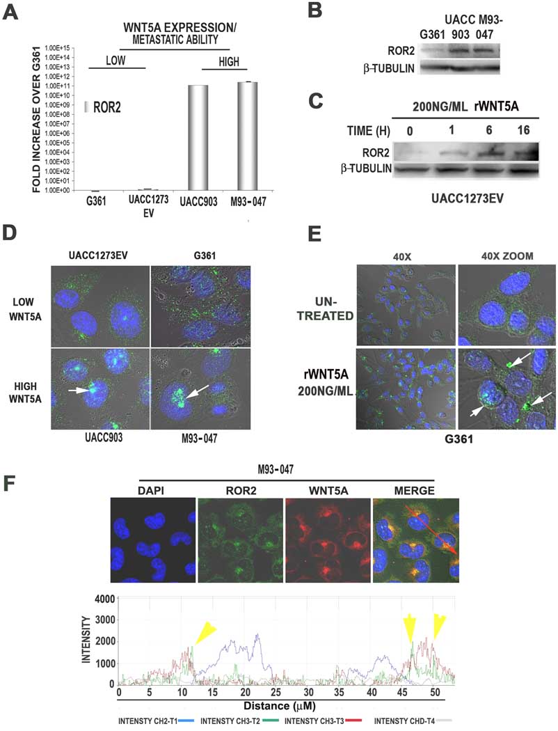
Figure 2. ROR2 expression and localization in human melanoma cell lines.
ROR2 mRNA is highly expressed in Wnt5A high lines (UACC903 and M93-047), but not Wnt5A low UACC1273EV and G361 cells (A). ROR2 protein levels are also higher in Wnt5A high lines than Wnt5A-low G361 cells, as shown by Western analysis (B). Treating ROR2-low UACC1273EV cells with rWnt5A increases ROR2 expression (C). Immunofluorescent analysis demonstrates that in Wnt5A-low lines, ROR2 is expressed in a diffuse cytoplasmic pattern in most cells. In Wnt5A-high lines, ROR2 is expressed in both a diffuse pattern as well as in perinuclear/ nuclear foci in the vast majority of the cells (D). Treatment of Wnt5A-low cells with rWnt5A increases the focal localization of ROR2 (E). Wnt5A and ROR2 expression are both expressed in the perinuclear foci, as shown by immunolocalization analysis (F).

