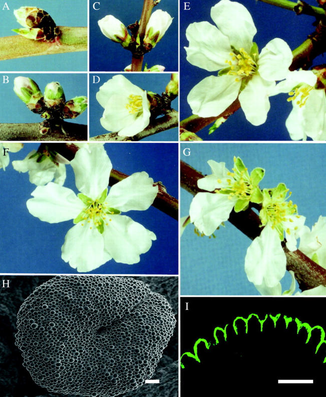
Fig. 1.

(A) Stage 1, swollen bud. (B) Stage 2, pink bud. (C) Stage 3, extended petals. (D) Stage 4, unfurling petals. (E) Stage 5, fully open. (F) Stage 6, flattened petals. (G) Stage 7, abscised petals. (H) SEM view of a stigmatic surface at stage 1. Scale bar = 80 µm. (I) Section through the surface of a stage 1 stigma stained with auramine O under fluorescence microscopy showing a cuticle layer on the surface. Scale bar = 40 µm.
