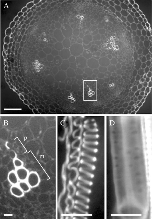
Fig. 1.

Xylem vessels in the basal region of inflorescence stems of arabidopsis observed under a fluorescence microscope. (A) Overview of a cross section of an inflorescence stem cut in the basal region. Scale bar = 100 µm. (B) Detail of xylem vessels in the area outlined in (A). m, Metaxylem; p, protoxylem. (C and D) Longitudinal view of the protoxylem (C) and the metaxylem (D) in a cleared stem. Scale bars = 10 µm.
