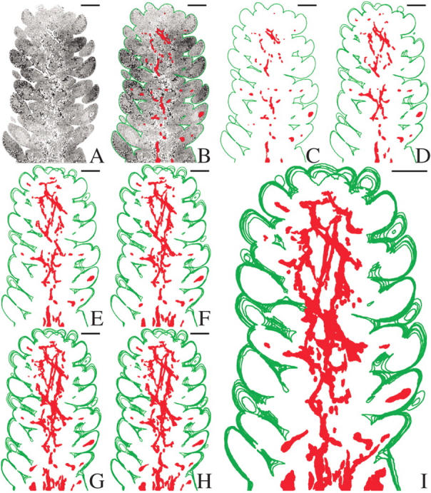
Fig. 1.

Stacked tracings of section profiles showing the distribution of the mistletoe endophyte in the cortical region near the periphery of a dormant Douglas fir apical bud. (A) Light micrograph of a longitudinal section of a dormant A. douglasii-infected Douglas fir apical bud. (B) Same as (A) but with host outlined in green and mistletoe cells highlighted in red. (C) Same as (A) and (B), but with all host cells masked to create a tracing representation of the mistletoe present in a single section. (D–I) Serial tracing representations, at 10-μm intervals, are stacked to illustrate the pattern of distribution of the endophyte in the cortex of the host. In (I) note the presence of the mistletoe in primordial leaves. Scale bars = 250 μm.
