Fig. 3.

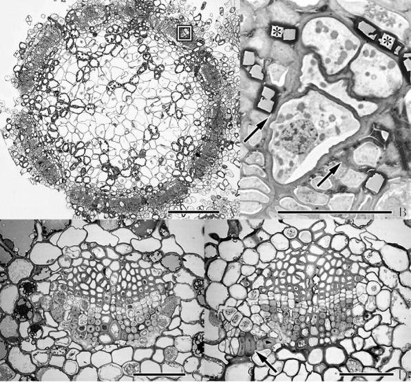
(A, B) Cross-section of branch and (C, D) needles of A. douglasii-infected Douglas fir. (A) SEM micrograph of cross-section through an infected branch proximal to the crown region of a dormant bud. (B) Close-up of box outlined in (A) in the region of a vascular bundle showing presumed sinker initiation by a multiseriate strand of mistletoe. Arrows indicate cambial zone. Primary xylem is at bottom of micrograph. Oxalic acid crystals are identified by asterisks. (C) Light micrograph of a cross-section of a vascular bundle of a healthy needle. (D) Same as (C) but with possible endophytic strand indicated by arrow near phloem. Scale bars: A = 250 μm; B–D = 50 μm.
