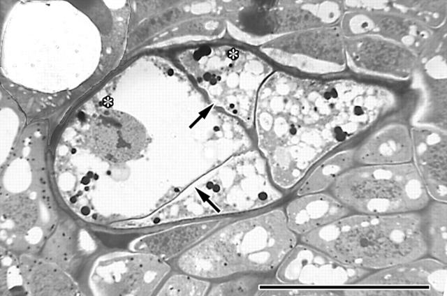
Fig. 4.
Light micrograph of cross-section of a multiseriate strand of the endophyte of A. douglasii infecting Pseudotsuga menziesii illustrating the five characteristics utilized to differentiate the parasite from its host: (1) thickened external endophyte cell walls; (2) cell size and shape; (3) chromocentric nucleus; (4) presence of lipids (asterisks); and (5) plasmolysis (arrows) and vacuolization. Scale bar = 50 μm.

