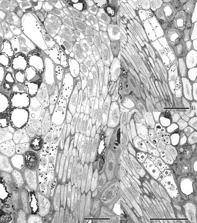
Fig. 5.

Light micrographs of endophytic filaments of A. douglasii near the crown region in the dormant apical buds of Douglas fir illustrating the tiered arrangement of cells characteristic of multiseriate strands. In each case, the endophytic filament is contiguous with and adjacent to the procambium. (A) Procambium is to the right of the endophyte in the lower two-thirds of the micrograph. This filament ultimately terminates in a leaf primordium. (B) Procambium is to the left of this clearly defined example of a multiseriate filament. (C) This micrograph demonstrates the ambiguity encountered in differentiating mistletoe from host. Only cells labelled with an asterisk were positively identified as endophyte. Scale bars = 50 μm.
