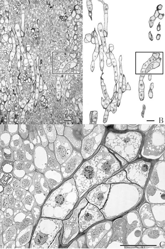
Fig. 6.

Light micrographs of a longitudinal section through the cortex of an infected dormant Douglas fir apical bud. (A) Unedited micrograph. (B) Same as (A), but with the host tissue hidden by an opaque mask. (C) Close-up of area outlined in (A) and (B) with the host partially masked to emphasize the two converged uniseriate strands of the endophyte. Scale bars = 50 μm.
