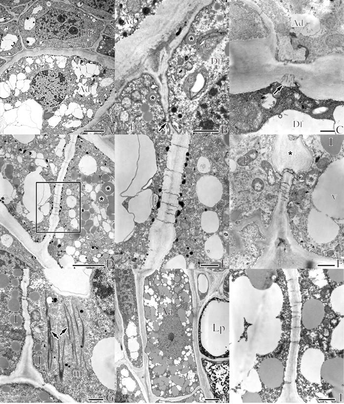
Fig. 8.

Transmission electron micrographs of A. douglasii (Ad) in dormant apical buds of Douglas fir (Df) (A–G) and of A. americanum (Aa) in the apical buds of lodgepole pine (Lp) (H and I). (A) Micrograph illustrating some of the most common ultrastructural features used to differentiate parasite from host, including the distinctive thick external cell wall, relatively larger cell size and increased vacuolization of the mistletoe cell (Ad) contrasted to the host cell (Df). Scale bar = 5 μm. (B) Close-up of the lower right corner of (A) illustrating some of the ultrastructural details of the host (Df), including chloroplasts with characteristic thylakoid membranes (c), lipids (asterisks) and plasmodesma (arrow). Scale bar = 1 μm. (C) Half plasmodesmata on the host side of the thickened external endophyte cell. Scale bar = 1 μm. (D) A portion of a uniseriate endophyte filament with characteristic thickened external cell wall, numerous lipids (asterisks) and plasmodesmata of the internal cell wall. Scale bar = 5 μm. (E) Area outlined in (D) illustrating plasmodesmata and small dark-stained starch granules. Scale bar = 1 μm. (F) Endophyte illustrating proplastids (p), lipids (l), vacuole (v) and an internal wall with numerous plasmodesmata and large cellulosic cell wall thickening (asterisk). Scale bar = 1 μm. (G) Ultrastructural details within a multiseriate strand of mistletoe with lipids (l), mitochondria (m) and endoplasmic reticulum (arrows). Scale bar = 1 μm. (H) A single cell of a uniseriate strand of the mistletoe endophyte; the cytoplasm is filled with numerous lipid bodies characteristic of late dormancy. Scale bar = 5 μm. (I) Close-up of an internal cell wall of the endophyte with the numerous plasmodesmata common to both of the Arceuthobium species studied. Scale bar = 1 μm.
