
Fig. 1.
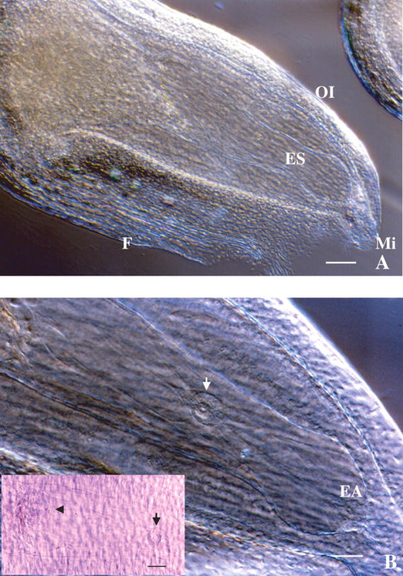
Microscopic observation of an Alstroemeria ovule using the clearing procedure. (A) Whole ovule showing the large embryo sac within. ES, Embryo sac; F, funicle; OI, outer integument; Mi, micropyle. Scale bar = 120 μm. (B) Magnification of the inside of an ovule. The egg apparatus (EA) and fused polar nuclei (white and black arrows) are in focus. The antipodal cells (arrowhead) are also observed in the left of the frame. Scale bar = 60 μm.