Figure 2.
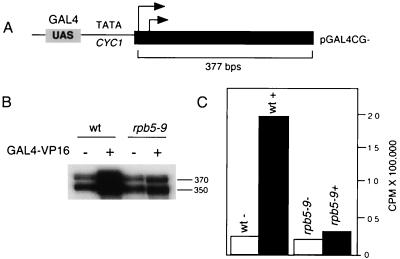
In vitro transcription of rpb5–9 reveals an activation defect. (A) DNA template used for transcription. Arrows represent approximate transcription start sites within the 377-bp G-less cassette. (B) Transcription products from wt and mutant (rpb5–9) whole-cell extracts with (+) or without (−) the addition of GAL4-VP16. mRNA sizes after gel electrophoresis and autoradiography are indicated. (C) PhosphorImager quantification of results shown in B.

