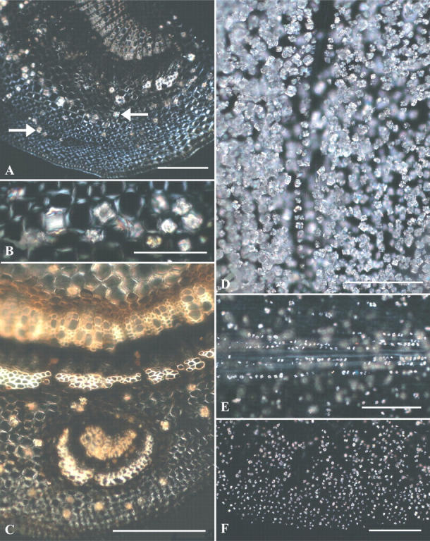
Fig. 3.

(A–F) Bright field (BF) and polarized light (PL) views of Prunus serotina stem sections and clearings of scales showing crystals. (A) Stem cross-section displays cortical druses (left-pointing arrow) and prisms (right-pointing arrow); PL. (B) Enlarged region of (A) displays both druses and prisms; PL. (C) Vascular leaf trace (below) in stem cross-section displays only druses in cortex; PL. (D) Thick outer bud scale filled with crystals; PL. (E) Inner bud scale showing midvein druses (in focus) and scattered druses elsewhere (out of focus); PL. (F) Thin inner bud scale margin with druses in all cell types; PL. Scale bars: A, C–F = 200 μm; B = 100 μm.
