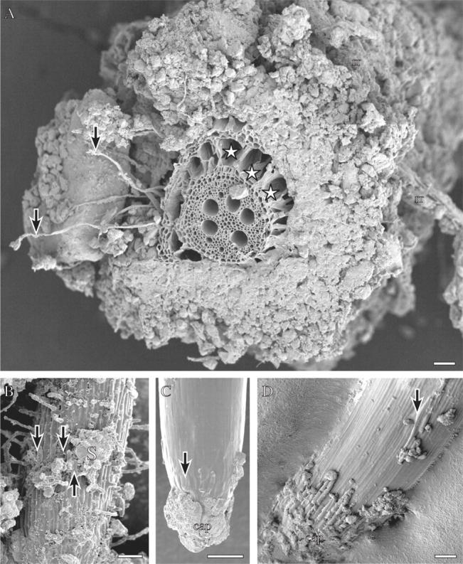
Fig. 1.

Wheat roots grown in the field (A, D), or in the same field soil in a rhizobox (B, C), frozen in liquid nitrogen and viewed frozen with a cryo-scanning electron microscope (see Watt et al., 2005, for details of method). (A) A nodal root sectioned by hand on a piece of wax with a single edged blade before freezing. Arrows indicate root hairs extending the length of the bound rhizosheath soil. A dead remnant root is caught in this soil (rr). Spaces (stars) have developed in the cortex. Scale bar = 300 µm. (B) Branch root with root hairs extending from the surface, bound soil (S) and root cap cells (arrows) along the surface. Scale bar = 100 µm. (C) Tip of the branch root in B. Root cap cells have collapsed (arrow) and soil adheres to the cap. Scale bar = 100 µm. (D) Tip of a nodal root. Sausage-shaped root cap cells are turgid on the cap and where sloughed along the elongation zone (arrow). Reproduced with permission from Watt et al. (2005). Scale bar = 200 µm.
