
Fig. 2.
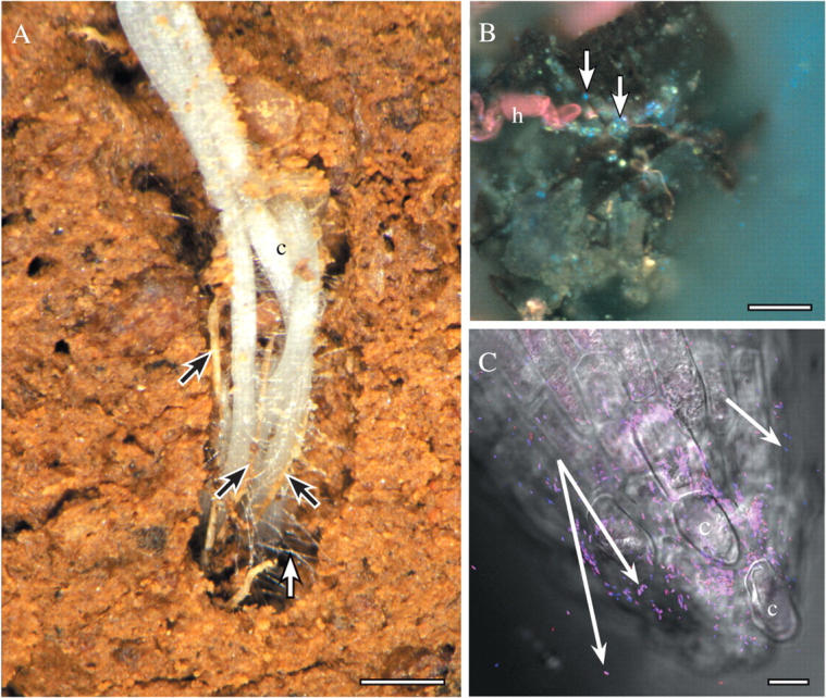
(A) Roots of canola (c) in the field, growing into a pore, in close contact with each other and dead roots of wheat (black arrows). Many root hairs (white arrow) extend from the new white, canola roots to bind to soil, and other living and dead remnant roots. Scale bar = 3 mm. (B) Root hair of wheat (h) associated with a piece of dark soil organic matter, bacteria (bright blue spots; some indicated by arrows) and mineral soil particles. Sample harvested from the field and prepared with the periodic-acid Schiff's reactions followed by DAPI (4,6 diamidine-phenyl indole), and viewed with UV fluorescence optics (see Watt et al., 2003). Scale bar = 20 μm. (C) Tip of wheat seminal root that was growing on agar and had Pseudomonas bacteria applied to the tip. Bacteria are hybridized to Bacteria- and Pseudomonas-specific oligonucleotide probes that are conjugated to fluorescent dyes, and viewed mounted in water, with a confocal microscope (reproduced from Watt et al., 2006a; see also for details of method). Some bacteria are bound to the root cap, and others are retained in hydrated mucilage behind the tip (white arrows). C, cap cell. Scale bar = 10 µm.