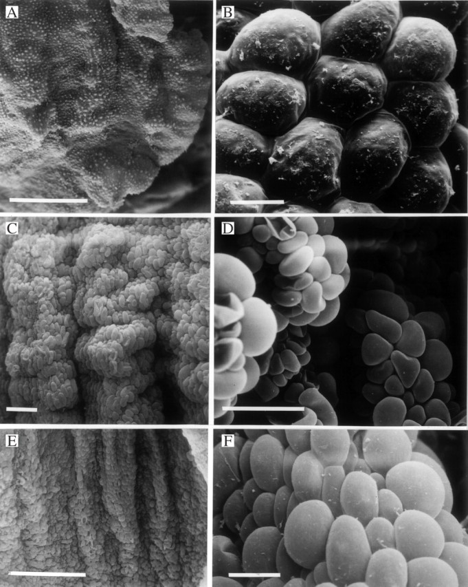
Fig. 5.

(A, B) Labellum of Xylobium corrugatum (K49592) (A) showing papillae with traces of secreted material (B). Scale bars: A = 500 μm; B = 10 μm. (C, D) Labellar surface of Xylobium bractescens (K14421) showing the arrangement (C) of laterally compressed, lollipop- or paddle-like papillae (D) along the carinae. Scale bars: C and D = 100 μm. (E, F) Labellum of Xylobium powellii (K8480) (E) showing laterally compressed papillae (F). Scale bars: E = 500 μm; F = 25 μm.
